

Can The Npnes In The Inner Ear Be Repaired
| Inner ear | |
|---|---|
 | |
| Details | |
| Artery | labyrinthine artery |
| Identifiers | |
| Latin | auris interna |
| MeSH | D007758 |
| TA98 | A15.3.03.001 |
| TA2 | 6935 |
| FMA | 60909 |
| Anatomical terminology [edit on Wikidata] | |

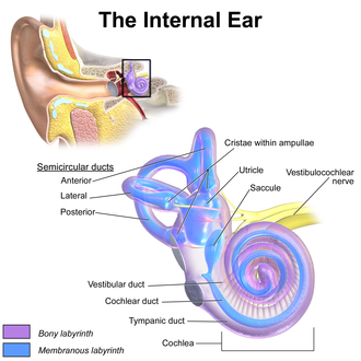
The inner ear (internal ear, auris interna) is the innermost part of the vertebrate ear. In vertebrates, the inner ear is mainly responsible for sound detection and balance.[1] In mammals, it consists of the bony labyrinth, a hollow cavity in the temporal bone of the skull with a system of passages comprising 2 principal functional parts:[two]
- The cochlea, defended to hearing; converting sound pressure patterns from the outer ear into electrochemical impulses which are passed on to the encephalon via the auditory nerve.
- The vestibular system, defended to residue
The inner ear is found in all vertebrates, with substantial variations in form and function. The inner ear is innervated past the 8th cranial nerve in all vertebrates.
Structure [edit]

The labyrinth can be divided by layer or by region.
Bony and membranous labyrinths [edit]
The bony labyrinth, or osseous labyrinth, is the network of passages with bony walls lined with periosteum. The three major parts of the bony labyrinth are the vestibule of the ear, the semicircular canals, and the cochlea. The membranous labyrinth runs inside of the bony labyrinth, and creates three parallel fluid filled spaces. The ii outer are filled with perilymph and the inner with endolymph.[3]
Vestibular and cochlear systems [edit]
In the middle ear, the energy of pressure level waves is translated into mechanical vibrations by the three auditory ossicles. Pressure waves move the tympanic membrane which in turns moves the malleus, the start bone of the middle ear. The malleus articulates to incus which connects to the stapes. The footplate of the stapes connects to the oval window, the kickoff of the inner ear. When the stapes presses on the oval window, it causes the perilymph, the liquid of the inner ear to motility. The middle ear thus serves to convert the energy from sound force per unit area waves to a forcefulness upon the perilymph of the inner ear. The oval window has only approximately 1/18 the area of the tympanic membrane and thus produces a college pressure. The cochlea propagates these mechanical signals as waves in the fluid and membranes and and then converts them to nerve impulses which are transmitted to the encephalon.[4]
The vestibular system is the region of the inner ear where the semicircular canals converge, close to the cochlea. The vestibular system works with the visual arrangement to proceed objects in view when the head is moved. Articulation and muscle receptors are also important in maintaining residual. The encephalon receives, interprets, and processes the information from all these systems to create the awareness of balance.
The vestibular arrangement of the inner ear is responsible for the sensations of balance and movement. It uses the same kinds of fluids and detection cells (pilus cells) equally the cochlea uses, and sends information to the brain about the attitude, rotation, and linear motion of the head. The type of motion or attitude detected by a hair cell depends on its associated mechanical structures, such as the curved tube of a semicircular canal or the calcium carbonate crystals (otolith) of the saccule and utricle.
Evolution [edit]
The human inner ear develops during week 4 of embryonic development from the auditory placode, a thickening of the ectoderm which gives ascent to the bipolar neurons of the cochlear and vestibular ganglions.[5] As the auditory placode invaginates towards the embryonic mesoderm, information technology forms the auditory vesicle or otocyst.
The auditory vesicle will give rise to the utricular and saccular components of the bleary labyrinth. They contain the sensory hair cells and otoliths of the macula of utricle and of the saccule, respectively, which respond to linear dispatch and the force of gravity. The utricular partitioning of the auditory vesicle also responds to angular dispatch, as well every bit the endolymphatic sac and duct that connect the saccule and utricle.
Beginning in the fifth week of development, the auditory vesicle too gives rise to the cochlear duct, which contains the spiral organ of Corti and the endolymph that accumulates in the bleary labyrinth.[6] The vestibular wall volition separate the cochlear duct from the perilymphatic scala vestibuli, a cavity inside the cochlea. The basilar membrane separates the cochlear duct from the scala tympani, a cavity within the cochlear labyrinth. The lateral wall of the cochlear duct is formed by the spiral ligament and the stria vascularis, which produces the endolymph. The hair cells develop from the lateral and medial ridges of the cochlear duct, which together with the tectorial membrane make up the organ of Corti.[vi]
Microanatomy [edit]


Cross-section through the spiral organ of Corti at greater magnification.
Rosenthal'south canal or the screw canal of the cochlea is a section of the bony labyrinth of the inner ear that is approximately 30 mm long and makes 2¾ turns about the modiolus, the central centrality of the cochlea that contains the spiral ganglion.
Specialized inner ear prison cell include: pilus cells, colonnade cells, Boettcher's cells, Claudius' cells, spiral ganglion neurons, and Deiters' cells (phalangeal cells).
The pilus cells are the master auditory receptor cells and they are too known as auditory sensory cells, acoustic pilus cells, auditory cells or cells of Corti. The organ of Corti is lined with a single row of inner hair cells and three rows of outer hair cells. The pilus cells accept a pilus bundle at the apical surface of the prison cell. The hair package consists of an assortment of actin-based stereocilia. Each stereocilium inserts equally a rootlet into a dense filamentous actin mesh known equally the cuticular plate. Disruption of these bundles results in hearing impairments and residue defects.
Inner and outer pillar cells in the organ of Corti support hair cells. Outer pillar cells are unique considering they are gratuitous standing cells which simply contact adjacent cells at the bases and apices. Both types of pillar cell have thousands of cantankerous linked microtubules and actin filaments in parallel orientation. They provide mechanical coupling between the basement membrane and the mechanoreceptors on the hair cells.
Boettcher'southward cells are found in the organ of Corti where they are present only in the lower plough of the cochlea. They lie on the basilar membrane beneath Claudius' cells and are organized in rows, the number of which varies betwixt species. The cells interdigitate with each other, and project microvilli into the intercellular space. They are supporting cells for the auditory hair cells in the organ of Corti. They are named subsequently German language pathologist Arthur Böttcher (1831-1889).
Claudius' cells are found in the organ of Corti located higher up rows of Boettcher's cells. Like Boettcher's cells, they are considered supporting cells for the auditory hair cells in the organ of Corti. They contain a multifariousness of aquaporin water channels and appear to be involved in ion send. They also play a function in sealing off endolymphatic spaces. They are named after the German anatomist Friedrich Matthias Claudius (1822-1869).
Deiters' cells (phalangeal cells) are a type of neuroglial prison cell found in the organ of Corti and organised in one row of inner phalangeal cells and three rows of outer phalangeal cells. They are the supporting cells of the pilus jail cell expanse within the cochlea. They are named later the German pathologist Otto Deiters (1834-1863) who described them.
Hensen's cells are high columnar cells that are direct adjacent to the third row of Deiters' cells.
Hensen's stripe is the section of the tectorial membrane to a higher place the inner hair prison cell.
Nuel'south spaces refer to the fluid-filled spaces between the outer colonnade cells and adjacent hair cells and as well the spaces between the outer hair cells.
Hardesty's membrane is the layer of the tectoria closest to the reticular lamina and overlying the outer hair cell region.
Reissner's membrane is composed of two jail cell layers and separates the scala media from the scala vestibuli.
Huschke's teeth are the tooth-shaped ridges on the spiral limbus that are in contact with the tectoria and separated past interdental cells.
Blood supply [edit]
The bony labyrinth receives its blood supply from three arteries: 1- Anterior tympanic branch (from maxillary artery). 2- Petrosal co-operative (from middle meningeal artery). 3- Stylomastoid branch (from posterior auricular artery). The membranous labyrinth is supplied by the labyrinthine artery. Venous drainage of the inner ear is through the labyrinthine vein, which empties into the sigmoid sinus or junior petrosal sinus.
Function [edit]
Neurons inside the ear reply to uncomplicated tones, and the brain serves to procedure other increasingly circuitous sounds. An boilerplate adult is typically able to detect sounds ranging betwixt 20 and twenty,000 Hz. The power to detect higher pitch sounds decreases in older humans.
The human ear has evolved with ii basic tools to encode sound waves; each is separate in detecting high and low-frequency sounds. Georg von Békésy (1899-1972) employed the use of a microscope in club to examine the basilar membrane located within the inner-ear of cadavers. He found that movement of the basilar membrane resembles that of a traveling wave; the shape of which varies based on the frequency of the pitch. In low-frequency sounds, the tip (apex) of the membrane moves the nearly, while in high-frequency sounds, the base of the membrane moves most.[7]
Disorders [edit]
Interference with or infection of the labyrinth can outcome in a syndrome of ailments called labyrinthitis. The symptoms of labyrinthitis include temporary nausea, disorientation, vertigo, and dizziness. Labyrinthitis can be acquired by viral infections, bacterial infections, or physical blockage of the inner ear.[8] [9]
Another condition has come to exist known every bit autoimmune inner ear disease (AIED). It is characterized by idiopathic, rapidly progressive, bilateral sensorineural hearing loss. It is a fairly rare disorder while at the same time, a lack of proper diagnostic testing has meant that its precise incidence cannot be determined.[10]
Other animals [edit]
Birds have an auditory organisation similar to that of mammals, including a cochlea. Reptiles, amphibians, and fish do not accept cochleas but hear with simpler auditory organs or vestibular organs, which generally detect lower-frequency sounds than the cochlea. The cochlea of birds is similar to that of crocodiles, consisting of a curt, slightly curved bony tube within which lies the basilar membrane with its sensory structures.[xi]
Cochlear system [edit]
In reptiles, audio is transmitted to the inner ear by the stapes (stirrup) bone of the middle ear. This is pressed against the oval window, a membrane-covered opening on the surface of the antechamber. From here, audio waves are conducted through a short perilymphatic duct to a second opening, the round window, which equalizes force per unit area, assuasive the incompressible fluid to move freely. Running parallel with the perilymphatic duct is a divide blind-ending duct, the lagena, filled with endolymph. The lagena is separated from the perilymphatic duct by a basilar membrane, and contains the sensory hair cells that finally translate the vibrations in the fluid into nerve signals. It is attached at one cease to the saccule.[12]
In nigh reptiles the perilymphatic duct and lagena are relatively short, and the sensory cells are bars to a small basilar papilla lying betwixt them. Withal, in mammals, birds, and crocodilians, these structures become much larger and somewhat more than complicated. In birds, crocodilians, and monotremes, the ducts are simply extended, together forming an elongated, more or less direct, tube. The endolymphatic duct is wrapped in a simple loop around the lagena, with the basilar membrane lying forth 1 side. The beginning one-half of the duct is now referred to equally the scala vestibuli, while the second half, which includes the basilar membrane, is chosen the scala tympani. As a result of this increase in length, the basilar membrane and papilla are both extended, with the latter developing into the organ of Corti, while the lagena is now called the cochlear duct. All of these structures together institute the cochlea.[12]
In therian mammals, the lagena is extended still further, condign a coiled structure (cochlea) in society to suit its length within the head. The organ of Corti besides has a more than complex structure in mammals than it does in other amniotes.[12]
The arrangement of the inner ear in living amphibians is, in most respects, like to that of reptiles. Nevertheless, they often lack a basilar papilla, having instead an entirely split up set of sensory cells at the upper edge of the saccule, referred to equally the papilla amphibiorum, which announced to accept the same role.[12]
Although many fish are capable of hearing, the lagena is, at best, a short diverticulum of the saccule, and appears to have no role in sensation of audio. Various clusters of pilus cells within the inner ear may instead be responsible; for case, bony fish contain a sensory cluster called the macula neglecta in the utricle that may have this function. Although fish have neither an outer nor a eye ear, sound may still be transmitted to the inner ear through the bones of the skull, or by the swim bladder, parts of which often lie close by in the body.[12]
Vestibular system [edit]
By comparison with the cochlear system, the vestibular system varies relatively trivial between the various groups of jawed vertebrates. The primal office of the system consists of two chambers, the saccule and utricle, each of which includes ane or two minor clusters of sensory hair cells. All jawed vertebrates too possess three semicircular canals arising from the utricle, each with an ampulla containing sensory cells at one end.[12]
An endolymphatic duct runs from the saccule up through the head and ending close to the brain. In cartilaginous fish, this duct actually opens onto the acme of the head, and in some teleosts, information technology is simply blind-ending. In all other species, nonetheless, it ends in an endolymphatic sac. In many reptiles, fish, and amphibians this sac may reach considerable size. In amphibians the sacs from either side may fuse into a single construction, which oft extends down the length of the body, parallel with the spinal canal.[12]
The archaic lampreys and hagfish, however, have a simpler arrangement. The inner ear in these species consists of a single vestibular chamber, although in lampreys, this is associated with a serial of sacs lined by cilia. Lampreys have only ii semicircular canals, with the horizontal culvert beingness absent, while hagfish take only a single, vertical, canal.[12]
Equilibrium [edit]
The inner ear is primarily responsible for balance, equilibrium and orientation in three-dimensional infinite. The inner ear can notice both static and dynamic equilibrium. Three semicircular ducts and ii chambers, which comprise the saccule and utricle, enable the body to detect whatever deviation from equilibrium. The macula sacculi detects vertical dispatch while the macula utriculi is responsible for horizontal acceleration. These microscopic structures possess stereocilia and one kinocilium which are located inside the gelatinous otolithic membrane. The membrane is further weighted with otoliths. Motion of the stereocilia and kinocilium enable the pilus cells of the saccula and utricle to find movement. The semicircular ducts are responsible for detecting rotational motion.[thirteen]
Boosted images [edit]
-
-

Ear labyrinth
-

Inner ear
-

Temporal bone
-

Right human bleary labyrinth, removed from its bony enclosure and viewed from the antero-lateral attribute
Run into besides [edit]
- Ear
- Hearing
- Middle ear
- Outer ear
- Tip link
References [edit]
- ^ Torres, M., Giráldez, F. (1998) The development of the vertebrate inner ear. Mechanisms of Development 71 (1-2) pg 5-21
- ^ J.Yard. Wolfe et al. (2009). Sensation & Perception. 2d ed. Sunderland: Sinauer Associated Inc
- ^ Rask-Andersen, Helge; Liu, Wei; Erixon, Elsa; Kinnefors, Anders; Pfaller, Kristian; Schrott-Fischer, Annelies; Glueckert, Rudolf (November 2012). "Human Cochlea: Anatomical Characteristics and their Relevance for Cochlear Implantation". The Anatomical Record: Advances in Integrative Anatomy and Evolutionary Biological science. 295 (xi): 1791–1811. doi:10.1002/ar.22599. PMID 23044521. S2CID 25472441.
- ^ Jan Schnupp, Israel Nelken and Andrew Male monarch (2011). Auditory Neuroscience. MIT Press. ISBN978-0-262-11318-2. Archived from the original on 2012-03-07. Retrieved 2011-04-13 .
- ^ Hyman, Libbie Henrietta (1992). Hyman'due south comparative vertebrate anatomy (3 ed.). University of Chicago Press. p. 634. ISBN0-226-87013-8 . Retrieved 2011-05-xiv .
- ^ a b Brauer, Philip R. (2003). Man embryology: the ultimate USMLE stride 1 review. Elsevier Wellness Sciences. p. 61. ISBNone-56053-561-X . Retrieved 2011-05-14 .
- ^ Schacter, Daniel (2012). Psychology. New York, NY: Worth Publishers. ISBN978-1464135606.
- ^ Labyrinthine dysfunction during diving. 1st Undersea and Hyperbaric Medical Lodge Workshop. Vol. UHMS Publication Number WS6-fifteen-74. Undersea and Hyperbaric Medical Order. 1973. p. xi. Retrieved 2009-03-11 .
- ^ Kennedy RS (March 1974). "General history of vestibular disorders in diving". Undersea Biomedical Inquiry. 1 (ane): 73–81. PMID 4619861. Retrieved 2009-03-11 .
- ^ Ruckenstein, K. J. (2004). "Autoimmune Inner Ear Disease". Current Opinion in Otolaryngology & Head and Neck Surgery, 12(five), pp. 426-430.
- ^ "Bird cochlea".
- ^ a b c d e f g h Romer, Alfred Sherwood; Parsons, Thomas S. (1977). The Vertebrate Torso. Philadelphia, PA: Holt-Saunders International. pp. 476–489. ISBN0-03-910284-10.
- ^ Anatomy & Physiology The Unity of Grade and Function. Northward.p.: McGraw-Colina Higher, 2011. Print.
- Ruckenstein, M. J. (2004). "Autoimmune Inner Ear Disease". Current Opinion in Otolaryngology & Caput and Neck Surgery, 12(v), pp. 426–430.
- Saladin, Anatomy and Physiology 6th ed., impress
- American Speech-Language-Hearing Clan, "The Middle Ear",
External links [edit]
| | Wikimedia Commons has media related to Inner ear. |
- Anatomy photograph:30:05-0101 at the SUNY Downstate Medical Center

Source: https://en.wikipedia.org/wiki/Inner_ear
Posted by: goldmanreaver.blogspot.com
0 Response to "Can The Npnes In The Inner Ear Be Repaired"
Post a Comment